Welcome to our depth exploration of the anatomy of the knee. In this detail guide, we will explore into the intricate details of the knee Anatomy, its components, functions, and common issues.
Whether you are a medical professional, a student, or simply curious about how your knee works, we will provide you with valuable insight and knowledge for knee anatomy.
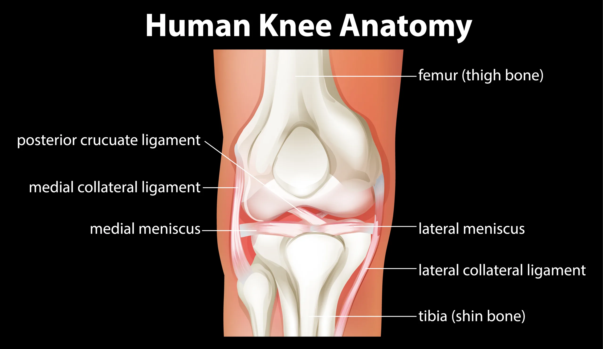
Basic Understanding the Knee Anatomy
The knee joint is a remarkable structure that plays an important role in supporting our body weight and facilitating movement. It is a complex hinge joint that connects the femur (thigh bone) to the tibia (shin bone).


The patella (kneecap) is another important component that articulates with the femur to form the patellofemoral joint.
Bones of the Knee Anatomy
Femur (thigh bone): The femur is the longest bone in the human body and forms the upper part of the knee joint.
Tibia (shin bone): The tibia is located below the femur and forms the lower part of the knee joint.
Patella (kneecap): The patella is a sesamoid bone that sits at the front of the knee joint, protecting it and exerting mechanical advantage.
Tendons and Ligaments
The knee joint is stabilized and supported by various ligaments and tendons, which play an important role in maintaining its integrity and function.

Anterior Cruciate Ligament (ACL): The ACL prevents forward movement of the tibia in relation to the femur and provides rotational stability.
Posterior Cruciate Ligament (PCL): The PCL prevents backward movement of the tibia in relation to the femur.
Medial Collateral Ligament (MCL): The MCL provides stability against forces that push the knee inward.
Lateral Collateral Ligament (LCL): The LCL provides stability against forces that push the knee outward.
Quadriceps tendon: This tendon attaches the quadriceps muscle to the patella and is essential for knee extension.
Patellar Tendon: The patellar tendon connects the patella to the tibia and aids in knee movements.

Functionality of the Knee
The knee joint is a classic example of biomechanics, allowing for a wide range of movements while maintaining stability and support. It enables vital activities such as walking, running, jumping and sitting.
Menisci
Menisci are crescent-shaped cartilages that are located between the femur and the tibia. They act as shock absorbers, distribute forces more evenly, and increase joint stability.
Synovial Fluid
The knee joint contains synovial fluid, which lubricates the joint, nourishes the cartilage, and reduces friction during motion.
Articular Cartilage
The surfaces of the femur, tibia and patella are covered with articular cartilage – a smooth, smooth tissue that reduces friction and absorbs shock during movement.
Common Knee Issues
Despite its complexity and flexibility, the knee anatomy is vulnerable to a variety of problems that can cause discomfort and hinder mobility.
Ligament Injuries
Injuries to the ligaments, such as an ACL tear, can result from a sudden twist or impact, causing instability and limited range of motion.
Meniscus Tears
Meniscus tears often occur during activities that involve bending the knee, causing pain, swelling, and limited range of motion.
Osteoarthritis
Osteoarthritis is a degenerative condition that affects articular cartilage, causing pain, stiffness, and decreased joint function.
Maintaining Healthy Knees
Taking care of your knees anatomy is essential to ensuring a healthy and active lifestyle. Here are some tips for knee care:
Stay active:
Engage in low-impact exercises to strengthen the muscles around the knee and improve joint stability.
Maintain a healthy weight:
Excess weight puts extra stress on the knee joint, increasing the risk of injuries and conditions like osteoarthritis.
Wear appropriate footwear:
Choose shoes that provide adequate support and cushioning to reduce stress on the knees.
Warm-up and cool down:
Always warm up before physical activity and do cool-down stretches to prevent muscle stiffness.
Use proper techniques:
Whether you are exercising or lifting objects, make sure you use proper techniques to avoid straining your knees.
Conclusion
Understanding the knee anatomy of the knee is important for maintaining optimal joint health and preventing potential issues. The complex design of the knee joint, including bones, ligaments, tendons and cartilage, enables us to perform a wide range of activities.
By taking a proactive approach to knee care and seeking medical guidance when needed, you can enjoy an active and fulfilling life while keeping your knees happy and healthy.
FAQ’s
Q1: What is knee joint?
The knee joint is a complex hinge joint that connects the thigh bone (femur) to the shin bone (tibia). It allows for flexion and straightening of the leg as well as some rotational movements.
Q2: What are the main components of the knee joint?
The knee joint is made up of the femur (thigh bone), tibia (shin bone) and patella (knee cap). It also includes various ligaments, tendons, muscles, and cartilage structures that provide stability and support.
Q3: What is the role of cartilage in the knee?
Cartilage in the knee, especially articular cartilage, acts as a cushion and reduces friction between the bones during motion. It helps to absorb shock and allows the joint surfaces to glide smoothly.
Q4: What are menisci?
The menisci are C-shaped pieces of cartilage located between the femur and the tibia. They increase stability, distribute weight and provide cushioning to the knee joint.
Q5: What are ligaments, and how do they function in the knee?
Ligaments are strong, flexible bands of connective tissue that help stabilize the knee joint. The major ligaments anterior cruciate ligament (ACL), posterior cruciate ligament (PCL), medial collateral ligament (MCL), and lateral collateral ligament .
Q6: What is the function of ACL and PCL?
The anterior cruciate ligament (ACL) prevents the tibia from moving too far forward relative to the femur, and the posterior cruciate ligament (PCL) prevents excessive backward movement of the tibia.
Q7: How do the MCL and LCL contribute to knee stability?
The medial collateral ligament stabilizes the inner part knee, while the lateral collateral ligament (LCL) stabilizes the outer part. They help prevent excessive side-to-side motion.
Q8: What is the function of synovial fluid in the knee joint?
Synovial fluid is a lubricating fluid that helps reduce friction within the knee joint. It nourishes cartilage and other structures while providing a smooth surface for walking.
Q9: What treatment options are available for knee injuries?
Treatment for knee injuries may include rest, physical therapy, medications, bracing, and in some cases, surgery. The choice of treatment depends on the nature and severity of the injury.
For More Blog On-Humanxplorer
Ear Infection Symptoms and Treatments-A Complete Guide
Weak Knees Causes and Treatments
What is a Cardiovascular Perfusionist-Heroes of Heart Surgery


3 thoughts on “Knee Anatomy: Causes, Treatments, Prevention-2023”